Lymph Node Identification
Computer Vision–Based Identification of Lymph Nodes in Surgical and Endoscopic Videos
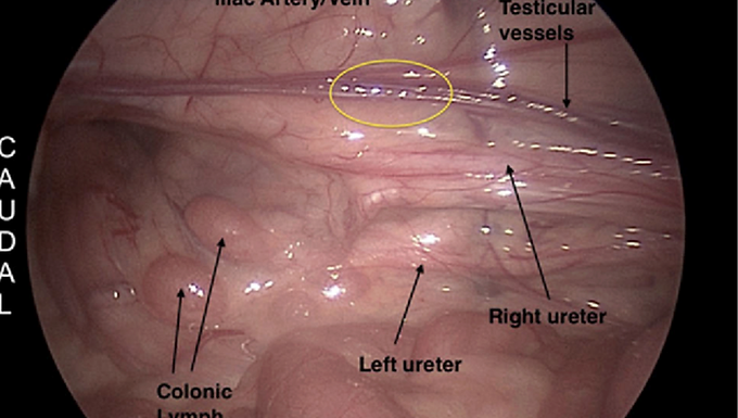
Accurate identification of lymph nodes during surgical and endoscopic procedures is critical for cancer staging, oncologic resection, and biopsy guidance. In many minimally invasive surgeries, lymph nodes are small, partially obscured, and visually similar to surrounding fat and connective tissue, making intra-operative identification challenging and highly dependent on surgeon experience.
The aim of this project is to develop a computer vision system that analyzes laparoscopic or endoscopic video to detect, localize, or segment lymph nodes in real time or offline analysis. Using real surgical video data, students will design, implement, and evaluate vision-based methods that highlight candidate lymph nodes and support surgical decision-making.
Mentor Details:
Prof. Yoav Mintz
Mentor Details:
Requirments:
Students will aim to:
Analyze surgical/endoscopic video data to characterize visual features of lymph nodes
Develop a computer vision pipeline for lymph node detection or segmentation
Apply deep learning–based models (e.g., CNNs, Vision Transformers) to image or video data
Incorporate temporal consistency to reduce false positives
Evaluate system performance using appropriate computer vision metrics
Problem Statement
Surgical and endoscopic videos pose significant challenges for automated lymph node identification:
Visual similarity between lymph nodes and surrounding adipose tissue
Variable node size, shape, and appearance
Occlusion by instruments, fat, or bleeding
Dynamic deformation and camera motion
Limited or noisy annotations due to clinical constraints
Unlike organs with well-defined boundaries, lymph nodes often appear as subtle, context-dependent structures, making reliable detection particularly difficult. The problem is to design a vision-based system that can consistently identify lymph nodes across frames or video sequences under real-world surgical conditions.
Project Objectives
Students will aim to:
Analyze surgical/endoscopic video data to characterize visual features of lymph nodes
Develop a computer vision pipeline for lymph node detection or segmentation
Apply deep learning–based models (e.g., CNNs, Vision Transformers) to image or video data
Incorporate temporal consistency to reduce false positives
Evaluate system performance using appropriate computer vision metrics
Technical Scope
The project may include one or more of the following components:
Object detection of lymph nodes in laparoscopic or endoscopic frames
Semantic or instance segmentation of lymph nodes
Multi-class classification (lymph node vs fat vs vessel)
Temporal tracking of detected nodes across video frames
Weakly supervised learning using surgical reports or biopsy confirmation
Required Knowledge and Prerequisites
Core Requirements
Familiarity with fundamental computer vision concepts
Experience with convolutional neural networks (CNNs)
Basic understanding of deep learning frameworks (e.g., PyTorch, TensorFlow)
Ability to work with image and video datasets
Recommended Background
Object detection and segmentation architectures (e.g., YOLO, Mask R-CNN)
Video processing and tracking techniques
Model evaluation metrics (precision, recall, F1-score, IoU)
No prior oncologic or surgical knowledge is required; relevant clinical context will be provided.
Project Difficulty and Expected Level
Vision complexity: High (small targets, low contrast, cluttered scenes)
Modeling complexity: Moderate to high
Domain knowledge: Low
This project is well-suited for:
Teams of 2–4 students
Expected Outcomes
A working computer vision prototype for lymph node identification
Quantitative evaluation on surgical or endoscopic video datasets
Analysis of false positives and missed detections
Well-documented code and a technical report